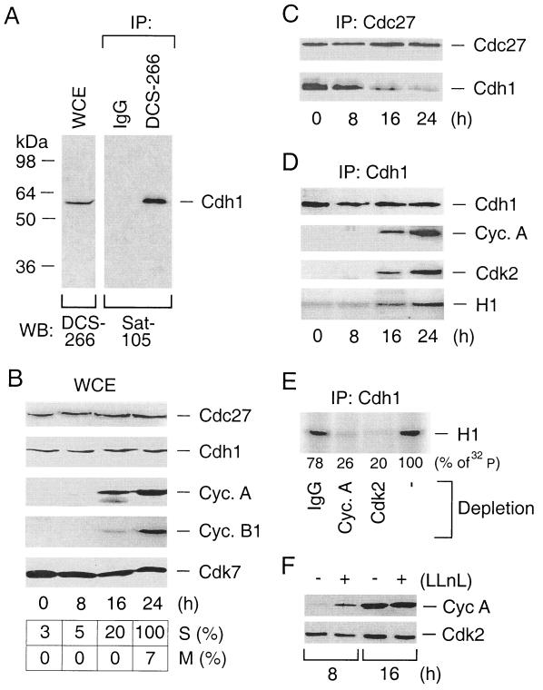
FIG. 1.
Endogenous Cdh1 redistributes from the APC to active cyclin A-Cdk2 complexes during the cell cycle. (A) Characterization of the DCS-266 mouse monoclonal antibody to Cdh1. Whole-cell extracts (WCE) prepared from R12 cells were either directly immunoblotted with DCS-266 (left) or immunoprecipitated (IP) with DCS-266 (right). DCS-266 immunoprecipitates were subjected to Western blotting analysis by Sat-105, an affinity-purified rabbit polyclonal antibody to Cdh1. IgG, immunoglobulin G. (B) R12 cells were starved for 48 h by incubating them in a mitogen-free medium and then induced to reenter the cell cycle by addition of FCS. At the indicated time points, the kinetics of cell cycle progression were measured by scoring BrdU-positive cells as an indication of productive entry into S phase (S) and by counting cells with condensed chromosomes as an indication of entry into mitosis (M). Expression profiles of Cdc27, Cdh1, cyclin A, cyclin B1, and Cdk7 (loading control) were assessed by Western blotting analysis of the WCE. (C) R12 cells were synchronized as for panel B. The APC was immunoprecipitated by an antibody to its Cdc27 structural subunit and analyzed for the presence of the Cdh1-activating subunit by Western blotting with Cdh1-specific antibody DCS-266. (D) R12 cells were synchronized as for panel B. Cdh1 complexes immunopurified by the DCS-266 antibody were analyzed for the presence of cyclin A and Cdk2 by Western blotting and for the associated kinase activity by an in vitro kinase assay using histone H1 as a substrate. (E) R12 cells were synchronized in early S phase by mitogen depletion for 48 h and subsequent restimulation by addition of FCS for 16 h. Cell extracts were immunodepleted with antibodies to cyclin A, Cdk2, or control IgG as indicated, and Cdh1 immunoprecipitates were subjected to an in vitro kinase reaction as for panel D. (F) R12 cells were starved as for panel B and stimulated for either 8 or 16 h by FCS to allow progression into G1 and early S phases, respectively. For the last 2 h, at each time point, the culture medium was supplied with LLnL. The cell extracts were analyzed for the abundance of cyclin A and Cdk2 (here serves as a loading control) by Western blotting.

