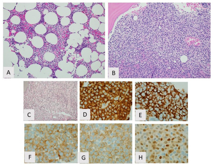
Figure 2.
Hairy cell leukemia, bone marrow. (A) Partial involvement showing typical interstitial growth pattern with “fried egg” appearance, H&E, 20×; (B) Diffuse involvement with sheet-like growth pattern H&E, 20×; (C) Increased reticulin fibrosis, reticulin stain, 20×. By immunohistochemistry the cells are positive for (D) CD20, (E) CD103 (F) CD25, (G) annexin A1, and (H) cyclin D1.

