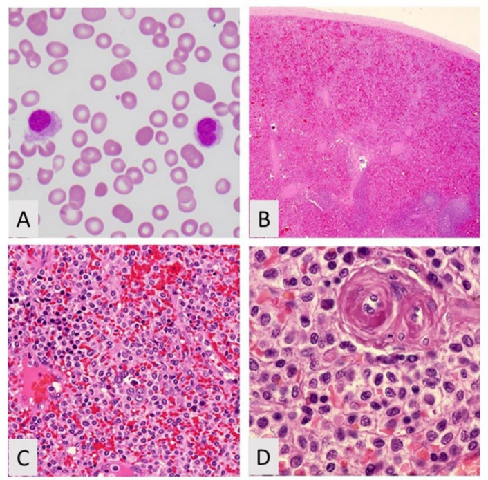
Figure 3.
Hairy cell leukemia. (A) Peripheral blood showing hairy cell, Wright–Giemsa, 400×; (B) Low power of spleen showing blood lakes H&R, 20×; (C) Intermediate power showing hairy cell leukemia and nucleated red blood cells, H&E, 200×; (D) Hairy cell leukemia showing cells with abundant cytoplasm, H&E, 400×.

