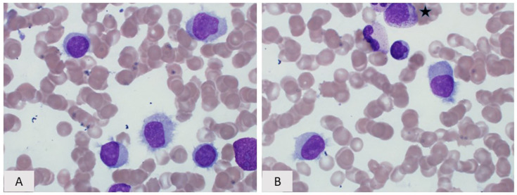
Figure 4.
Hairy cell leukemia variant. (A) Peripheral blood smear and (B) Bone marrow aspirate, (a myelocyte is present in the top of the field ★), Wright–Giemsa, 400×. The cytologic features range from typical “hairy cells” showing prominent circumferential cytoplasmic projections, rounded nuclei, spongy chromatin, and inconspicuous/absent nucleoli to cells with less prominent cytoplasmic projections, more finely dispersed chromatin m, and occasional prominent nucleoli.

