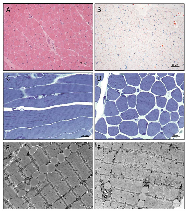
Figure 3.
Results of histological and electron microscopic studies of quadriceps muscle derived from our SLC18A3 patient. (A) Widened spectrum of muscle fibre calibres identified by H&E staining. (B) Increased number and size of lipid droplets in many muscle fibres identified by Oil Red O histochemistry. Scale bars = 50 µm. (C,D) Semithin resin section histology confirms the considerable widening of the calibre spectrum combined accumulation of osmiophilic material in many muscle fibres. Scale bars = 20 µm. Electron microscopic studies revealed (E) increased number of intermyobrillar lipid droplets, often arranged in rows and (F) minor focal Z-band disintegration. Scale bars = 2 µm.

