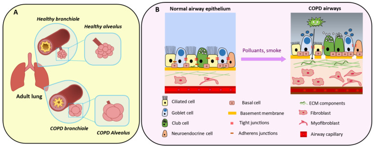
Figure 5.
Aspects of COPD development. (A) (Top panel) Healthy bronchioles present a thin wall and a lumen where mucus is secreted to externalize pathogens. Alveoli contain elastin fibers and regulate gas exchanges between lung and blood. (Bottom panel) Over time, chronic exposure to tobacco, air pollution and/or recurrent infections lead to a thickened bronchiole wall associated with airflow obstruction. Emphysema is caused by gas trapping that damages the alveoli, followed by progressive peribronchiolar fibrosis. (B) (Right panel) In the alveolar-capillary unit, epithelial cells are inter-connected and attached to the basement membrane above the mesenchymal compartment. (Left panel) Chronic exposure to pollutants damages ciliated cells (leading to impaired mucociliary clearance), alters the epithelial cell barrier and the basement membrane with loss of tight junctions and pathogen penetration in the lower layers. This is associated with mucus plugging and goblet cell hypersecretion. Pro-proliferative fibroblasts and myofibroblasts contribute to extracellular matrix (ECM) deposition that impairs injury repair.

