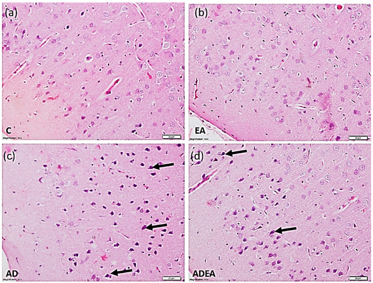
Figure 3.
Photomicrograph showing the normal architectural pattern of the ERC with neurons having vesicular pale nuclei in the control (a) and EA (b) groups. In the AD (c) group, the ERC exhibited disturbed architecture, and neurons with condensed deeply stained pyknotic nuclei (arrows). In the ADEA (d) group, most neurons were restored with clear vesicular nuclei among a few scattered hyperchromatic condensed nuclei (arrows). (H&E, magnification ×40, scale bar 20 µm).

