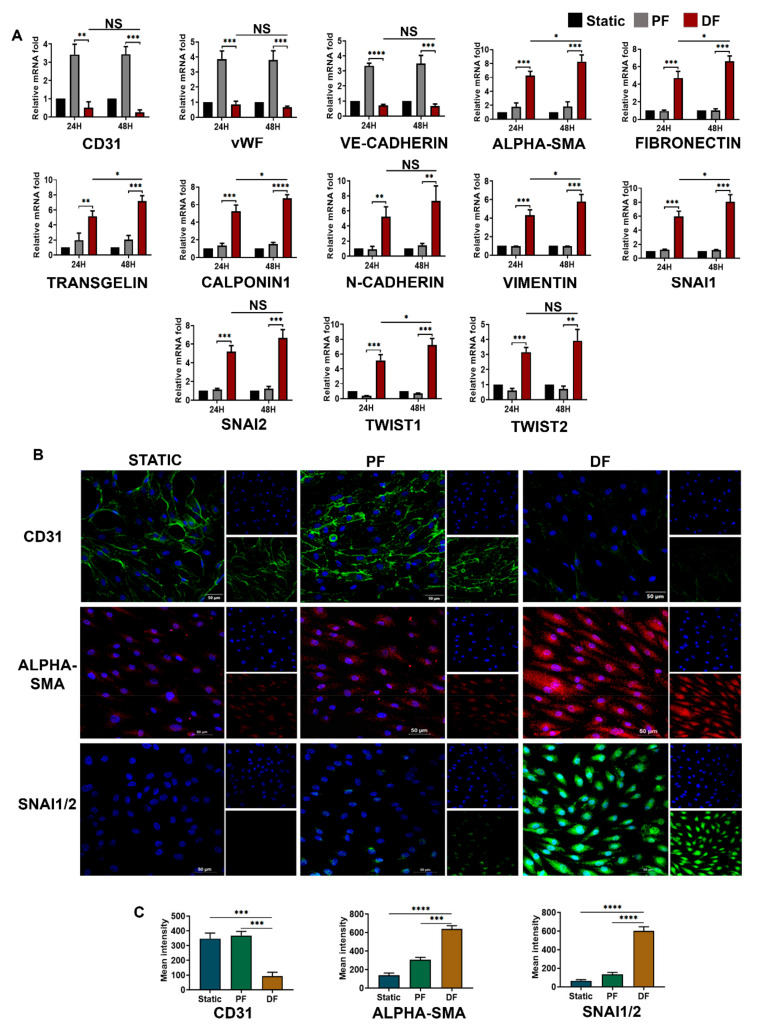
Figure 3.
EndMT activation by disturbed fluid flow in venous endothelial cells. (A) Time-dependent (24 and 48 h) mRNA-level expression of EndMT-associated transcriptional factors, endothelial markers, and mesenchymal markers upon exposure of HUVEC to disturbed flow (n = 3). EndMT becomes prominent as endothelial cells are exposed to disturbed flow for longer periods. The loss of endothelial markers and gain of mesenchymal markers was significant after exposure to disturbed flow for 48 h. mRNA fold values in parallel and disturbed flow were calculated relative to the static control. All data were normalized with GAPDH expression and are given as relative to static control. (B) HUVECS exposed to disturbed flow at 6 dyn/cm2 for 24 h resulted in an intermittent pattern of membrane-bound CD31 expression with an intense α-SMA staining compared to parallel uniform flow and static control conditions. SNAI1/2 transcription factors expression was augmented in the cells exposed to disturbed flow. SNAI1/2 expression was mostly confined to the cell nucleus, but several cells had both nuclear and cytoplasmic SNAI1/2 localization too (scale bar 50 µM, magnification 40×). (C) Mean fluorescence intensity was plotted as the average fluorescence intensity ± standard deviation (SD) of five microscopic fields per flow condition and from three biological replicates. * represents p < 0.05, ** p < 0.01, *** p < 0.001, and **** p < 0.0001 vs. respective static or parallel uniform shear-treated groups.

