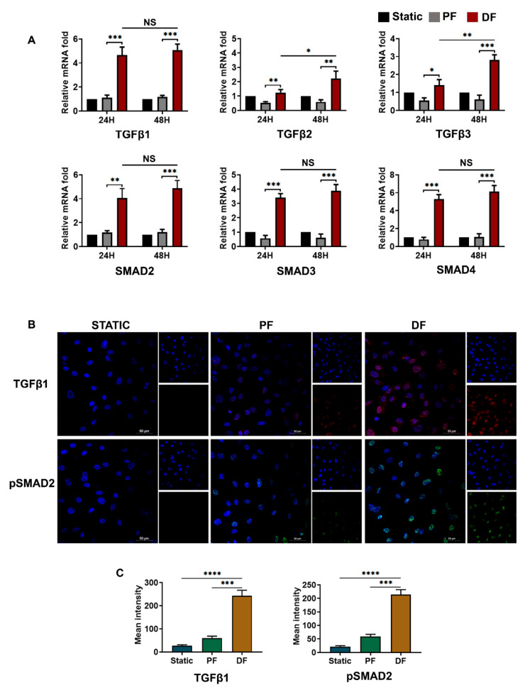
Figure 4.
TGFβ activation by disturbed fluid flow in venous endothelial cells. (A) Time-dependent (24 and 48 h) mRNA expression of TGFβ1, TGFβ2, and TGFβ3 ligands as well as their signaling partners SMAD2, SMAD3, and co-SMAD4 upon exposure of HUVEC to disturbed flow (n = 3). TGFβ1 and SMADs showed significant differences in expression between endothelial cells exposed to disturbed flow and parallel uniform flow as early as 24 h of exposure. The expression of TGFβ2 and TGFβ3 was significant after exposure to disturbed flow for 48 h. mRNA fold values were calculated relative to static control. All data were normalized with GAPDH expression and are given as relative to static control. (B) HUVECS exposed to disturbed flow at 6 dyn/cm2 for 24 h, showed overexpression of TGFβ1 and pSMAD2 (scale bar 50 µM, magnification 40×). (C) Mean fluorescence intensity was plotted as the average fluorescence intensity ± SD of five fields per flow condition and from three biological replicates. Analysis of five fields indicated an active TGFβ1- pSMAD2 pathway in the cells exposed to disturbed flow. PF indicates parallel uniform shear stress, and DF represents disturbed shear stress without any oscillatory flow. * indicates p < 0.05, ** p < 0.01, *** p < 0.001, and **** p < 0.0001 vs. respective static or parallel uniform shear-treated groups.

