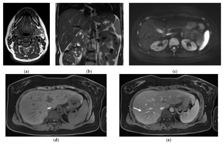
Figure 1.
Images illustrating the 20-min-WB-MRI-protocol: example of axial T1-weighted (T1w) TSE Dixon of the neck (a), coronal T2-weighted (T2w) HASTE of the upper abdomen (b), simultaneous multislice diffusion-weighted imaging of the abdomen and pelvis (c), and axial pre- (d) and post-contrast T1w VIBE Dixon from thorax to pelvis (e) in a 36-year old patient with malignant melanoma and currently no evidence of disease. Note that the sharpness of the anatomic structures, such as the liver vessels (arrow), was rated as excellent. Please note that the images have been cropped, as the skin tissue was part of the acquisition.

