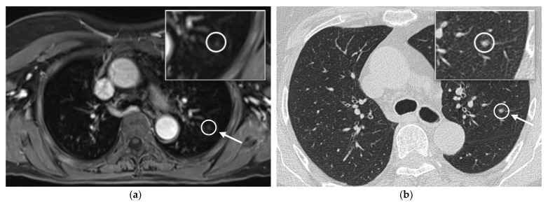
Figure 3.
Example of the assessment of the lung in the 20-min-WB-MRI-protocol and the recommended further CT-scan: example of axial post-contrast T1w VIBE Dixon of the thorax (a) and CT-scan one week later (b) in a 73-year old patient with malignant melanoma and currently no evidence of disease. In the WB-MRI scan, a new pulmonary nodule (arrow) was detected and a further CT scan was recommended. Please note that the images have been cropped, as the skin tissue was part of the acquisition.

