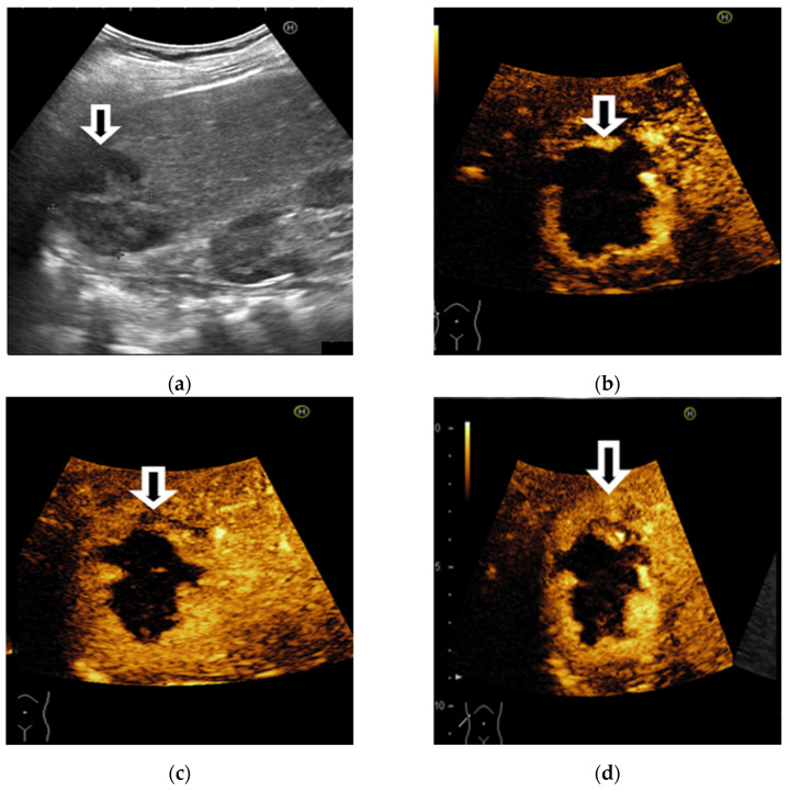
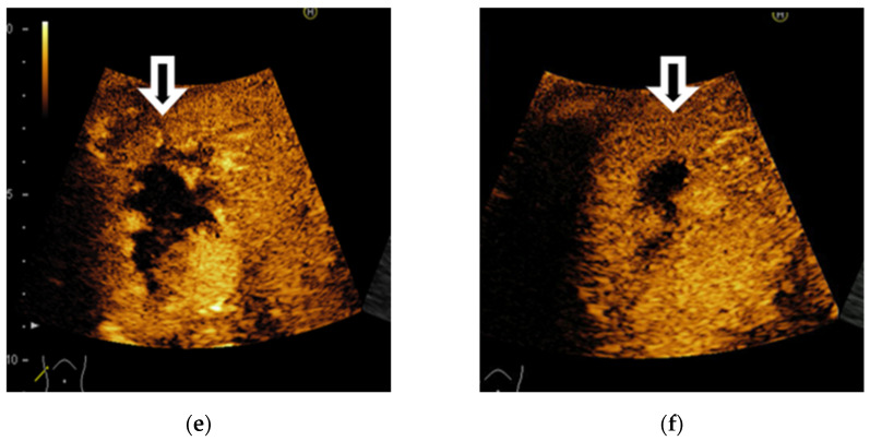
Figure 5.
A case of focal liver lesions detected in a 68-year-old man with liver cirrhosis, characterised as LI-RADS-1 on CEUS. On B mode ultrasound is observed a hyperechoic inhomogeneous liver, nodular liver surface and a hypoechoic, inhomogeneous FLL in the right liver lobe (a). On CEUS, the liver lesion shows a typical early, peripheral, globular enhancement (b,c) and centripetal fill-in (d,e). In the late phase, incomplete enhancement is noticed (f).


