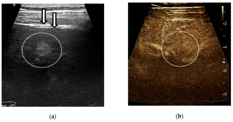
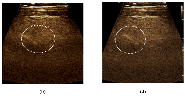
Figure 6.
Example of LI-RADS-2 liver lesion. Regenerative nodule <10 mm depicted by linear probe exam in a patient with hepatitis C cirrhosis. The ultrasound exam shows a nodular liver surface and a subcapsular hyperechoic liver lesion (a). On contrast-enhanced ultrasound (CEUS) the nodule shows the isoenhancing aspect in the arterial (b), portal-venous (c) and late phase (d).


