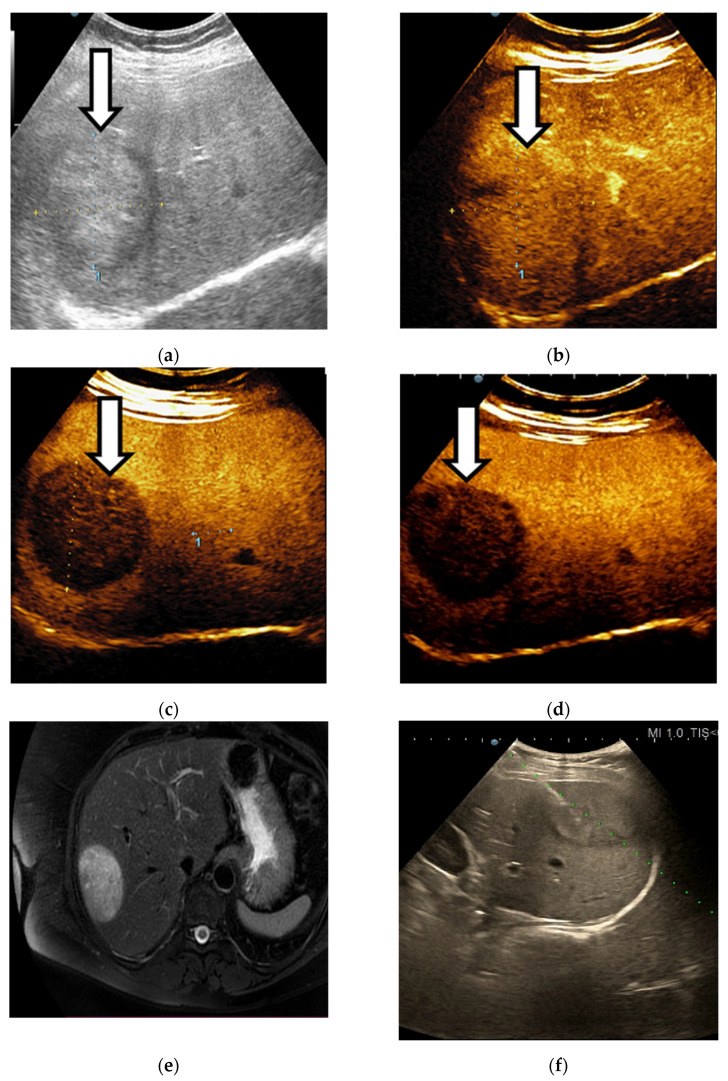
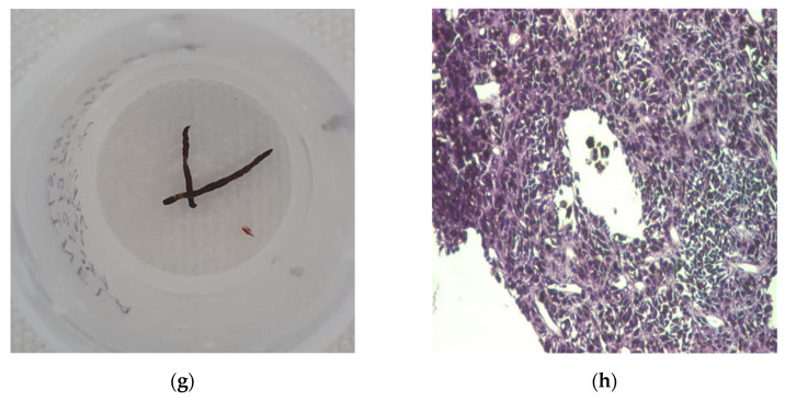
Figure 9.
A case of liver metastases detected in a 64-year-old woman with chronic hepatitis B, LI-RADS-M aspect. The ultrasound exam shows a hyperechoic lesion with a halo in the right liver lobe (a). On CEUS, the liver lesion shows an arterial phase enhancement (b). In the portal phase an early washout was noticed (c), followed by marked washout in the late phase (d). The aspect of focal liver lesion on the CT scan (e). Echo-guided liver biopsy was performed (f). Two black liver fragments (g) with melanoma histological aspect were obtained (×40) (h).


