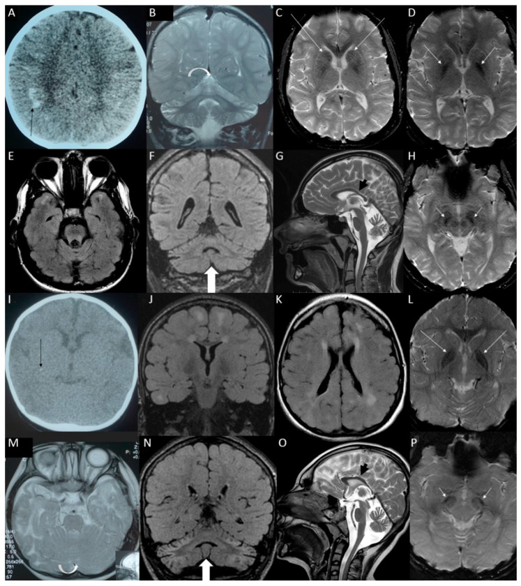
Figure 1.
Brain imaging of sibling with SUPV3L1 homozygous variant c.1093C>T:p.(Arg365Trp)—the older brother (patient S19a) in panels. (A) Computed tomography (CT) at the early childhood with marked subcortical calcifications in both frontal and parietal lobes (black arrow). (B) Subsequent magnetic resonance imaging (MRI) scans at the middle childhood with marked shrunken bright cerebellum (white arrow). (C–H) Brain MRI at the late adolescence. (E) FLAIR hyperintensities remained in the temporal poles only, (F) atrophic cerebellum but not so FLAIR-hyperintense as earlier and as in the younger sister (white arrow), (G) the corpus callosum is shorter and thinner than normal (black arrow), (D) iron or calcium deposits in the globi pallidi (white arrows), (H) in substantia nigra (white arrows) (C) and in the caudate nuclei (white arrows); brain imaging of sibling with SUPV3L1 homozygous variant c.1093C>T:p.(Arg365Trp)—the younger sister (patient S19b): (I) CT at the early childhood revealed two punctate subcortical calcifications in the right cerebral hemisphere (black arrow). (M) MRI taken as the toddler with normal cerebellum with slight widening of the cerebellar sulci (white arrow). (J–P) MRI at the early adolescence: (J,K) myelination has progressed, but there are white matter hyperintensities on FLAIR sequence in both cerebral hemispheres, (N) shrunken bright cerebellum (white arrow), (O) shortened and thinned corpus callosum (black arrow), (L) iron or calcium deposits in the globi pallidi (white arrows) (P) and in substantia nigra (white arrows).

