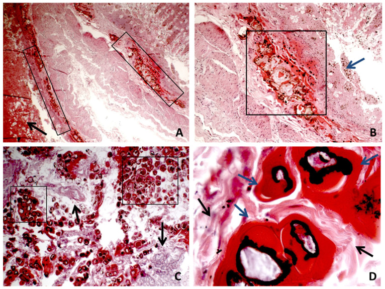
Figure 2.
(A,B) The panoramic of wall interruption, with hemorrhage, necrosis, inflammation, and Enterobius vermicularis accumulation in different bowel sections. The black arrow indicates large areas of bleeding. The black box shows the presence of cross-sections of EV with interruption of the muscolaris mucosa. The blue arrow indicates the mucosa that is no longer recognizable cytologically due to destruction and necrosis ((A) H&E ×4 magnification; (B) H&E ×10 magnification). (C) Area with a more evident presence of EV. The black box shows cross sections of EV; the black arrow indicates the residues of the intestinal wall fragmentation (H&E ×10 magnification). (D) High magnification of the transverse sections of the EV body; the black arrow indicates the residues of the intestinal wall fragmentation and the blue arrow indicate the EV body (H&E ×40).

