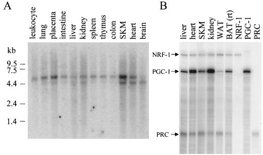
FIG. 3.
Expression profile of PRC mRNA. (A) A human multiple-tissue Northern blot (Clontech) probed with a 32P-labeled 1.6-kb PRC cDNA fragment comprising the 5′ end of the cDNA. Positions of RNA standards of known length in kilobases are indicated at the left. (B) Comparison of NRF-1, PGC-1, and PRC mRNA expression by RNase protection in various mouse tissues. Protected fragments (365, 297, and 192 bp for NRF-1, PGC-1, and PRC, respectively) from individual probes using liver RNA are shown in the last three lanes. WAT, white adipose tissue, rt, room temperature.

