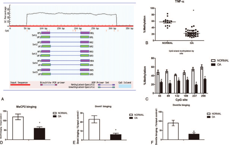
Figure 2.
DNA hypo-methylation in the TNF-α promoter in cartilage from OA patients. Figure 2: Methylation of the TNF-α locus in non-arthritic OA knee cartilage. Methylation was assessed by pyrosequencing of bisulphite converted genomic DNA. The CpG island spans from 50 to 320 bp upstream of the TNF-α TSS,which contains lots of CpG sites(A). Percentage overall methylation of the CpG island in cartilage from Normal (n = 13) and OA knee (n = 37) patients(B). Methylation of the TNF-α island by CpG site(C). ChIP was performed to measure MeCP2 (D), Dnmt1 (E) and Dnmt3a (F) binding to the TNF-α promoter in normal and OA cartilage. IgG was used as a negative control. The results are expressed as the percentage of MeCP2 and Dnmt1/3a binding in the input control. Data are shown as the means ± SD. ∗P < .05, OA cartilage vs. normal cartilage. The data were obtained from 37 OA patients receiving knee replacement surgery and 13 samples of non-arthritic tissues obtained during amputation.

