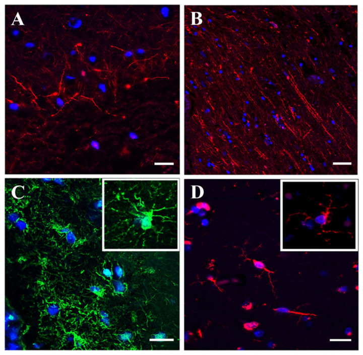
Figure 3.
Immunofluorescence analysis of human stroke brain tissue IF staining of human stroke brain tissue collected at 48 h after stroke onset. A, B: Pan-neuronal antibody staining (red) visualizing small unipolar and bipolar (A) and spindle-shaped (B) neurons. (C): GFAP (green) immunostaining for astrocytes. (D): Iba-1 (red) immunostaining for microglia. Bars: (A,C,D): 20 μm, and (B): 50 μm.

