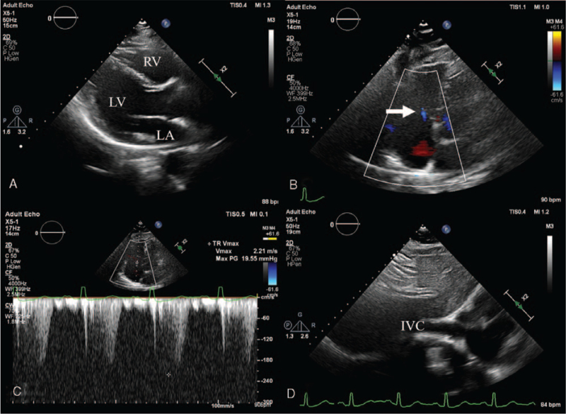
Figure 3.
Six-month follow-up echocardiographic images after attainment of euthyroid state showing (A) normalized size of the right ventricle, (B) near complete resolution of tricuspid regurgitation (arrow), and (C–D) normalization of pulmonary artery systolic pressure and inferior vena cava diameter. IVC = inferior vena cava, LA = left atrium, LV = left ventricle, RV = right ventricle.

