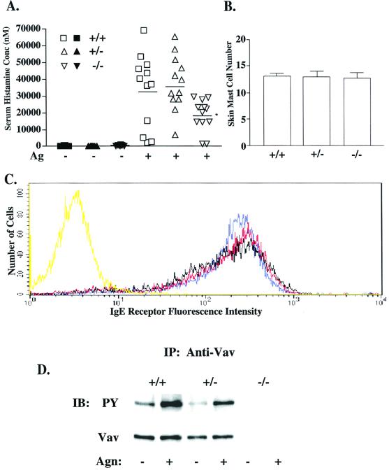
FIG. 1.
Effect of Vav1 deficiency on passive systemic anaphylaxis, IgE receptor expression, and Vav tyrosine phosphorylation. (A) Vav+/+ (squares), Vav+/− (triangles), and Vav−/− (inverted triangles) mice (n = 12 per dose group) were passively sensitized and challenged to induce systemic anaphylaxis as described in Materials and Methods. The serum histamine concentration (nanomolar concentration) was determined via inhibition enzyme-linked immunosorbent assay and compared among PBS (filled symbols)- and Ag (open symbols)-challenged animals, with the mean for each dose group indicated by a line. The asterisk indicates a significance of P ≤ 0.03 (t test) for Vav−/− responders compared to Vav+/+ mice. No significant difference was found between Vav+/+ and Vav+/− mice. (B) A section of depilated dorsal skin was removed from each animal (n = 2 per phenotype), and three sections per skin sample were stained with toluidine blue for mast cells. Mast cell number was assessed at a magnification of ×200 in random fields. (C) Vav+/+ (blue), Vav+/− (red), and Vav−/− (black) BMMC cultures (4 weeks) were sensitized with IgE and stained with an FITC-conjugated anti-IgE Ab to indirectly determine BMMC surface IgE receptor expression by flow cytometry. Nonspecific binding was determined by an FITC-conjugated isotype control (yellow). (D) Vav was immunoprecipitated from lysates of BMMCs (30 × 106) stimulated or not with 100 ng of Ag for 2 min, resolved by sodium dodecyl sulfate-polyacrylamide gel electrophoresis, and transferred to a nitrocellulose membrane. Tyrosine phosphorylation was determined by blotting with the antiphosphotyrosine Ab (PY, upper panel). After stripping, protein levels were determined by reblotting with anti-Vav Ab (lower panel). IP, immunoprecipitation; IB, immunoblotting.

