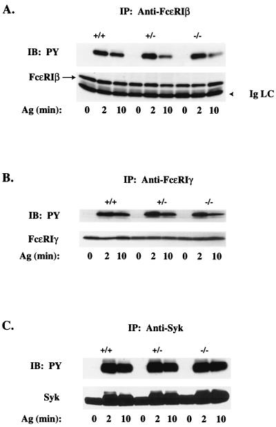
FIG. 3.
Tyrosine phosphorylation of the IgE receptor and Syk is Vav1 independent. Lysates were prepared from IgE-sensitized Vav+/+, Vav+/−, and Vav−/− BMMCs (30 × 106/ml) stimulated for 0, 2, or 10 min with 100 ng of Ag. FcɛRI β chain (A), FcɛRI γ chain (B), and Syk (C) were immunoprecipitated, resolved on sodium dodecyl sulfate-polyacrylamide gels, and transferred to nitrocellulose membranes. Tyrosine phosphorylation (PY) was determined by blotting with the antiphosphotyrosine Ab (upper panels). Individual protein levels were determined by reblotting with protein-specific Abs (lower panels). FcɛRI β protein is identified with an arrow to distinguish it from Ig light chain (IgLC). One representative of three experiments is shown. IP, immunoprecipitation; IB, immunoblotting.

