Figure 1.

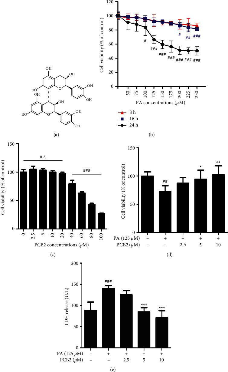
PCB2 protects HepG2 cells from PA-induced cell injury. (a) The chemical structure of PCB2. (b) HepG2 cells were treated with various concentrations of palmitic acid (from 50 to 250 μM) for 8, 16, and 24 h respectively. Cell viability was measured using CCK-8 and normalized to control (%). (c) Cytotoxicity of PCB2 (from 2.5 to 100 μM) for 24 h. (d) HepG2 cells were exposed to PA (125 μM) and treated by PCB2 (2.5, 5, and 10 μM) for 24 h. (c) HepG2 cells were treated as in (d), and (e) LDH released in the supernatant of cells was measured with a detection kit. The data are presented as means ± S.D. ##P < 0.01 and ###P < 0.001 vs control, ∗P < 0.05, ∗∗P < 0.01, and ∗∗∗P < 0.001 vs model group.
