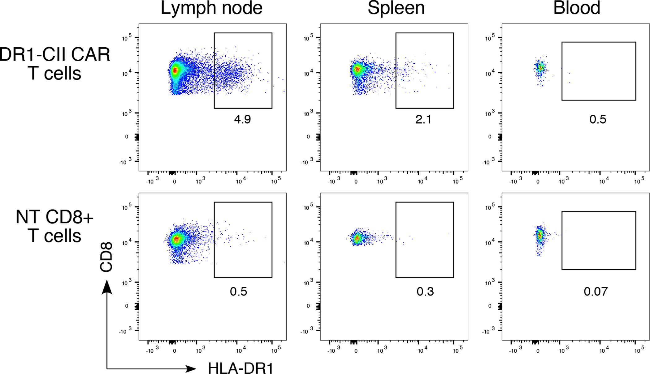
Figure 7. Detection of CAR T cells in lymphoid tissue 4 days after adoptive transfer.

B6.DR1 mice were immunized with CII/CFA, and 7 days later, mice were treated with 2 × 10⁶ DR1-CII CAR T-cells or NT CD8+ T cells (negative control). Four days after treatment, cells from draining lymph nodes, peripheral blood, and spleens were collected, incubated with fluorochrome labeled antibodies specific for CD3, CD19, CD4, CD8, and DR, stained with DAPI, and analyzed by flow cytometry. Data shown were generated from a single mouse and are representative of 3 experiments. Flow data were gated on CD3+/CD19−, and CD4−/CD8+, and are based on a minimum of 10,000 cells analyzed.
