
Figure 1: Anti-CD47 does affect atherogenesis or efferocytosis in the absence of LRP1 in vivo.
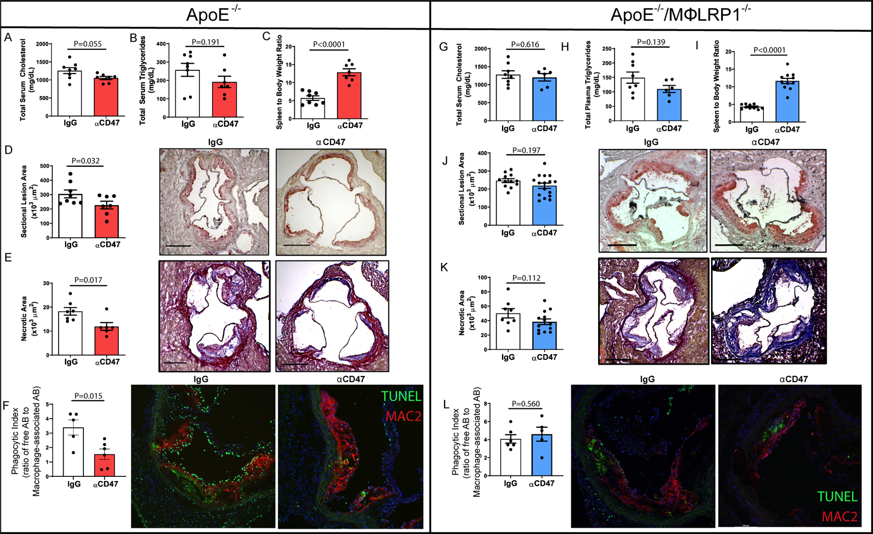
ApoE−/− mice were fed Western-type diet for 12 weeks while receiving anti-CD47 antibody (αCD47; 200μg/injection; N=7) or IgG (N=8) every other day. Mice were bled prior to euthanasia and serum was collected to determine A) Total serum cholesterol and B) total serum triglycerides. C) Spleen to body weight ratios were reported. Hearts were used to obtain frozen serial sections of aortic sinus. Frozen sinus sections were stained with D) Oil red-O to quantify lesion area (x103 μm2) One-tailed Student’s t-test was performed to determine differences between treatments. *P<0.05. E) Masson’s Trichrome stain was performed, and necrotic core area was quantified as regions of acellularity (x103 μm2). F) Immunofluorescent staining was performed using antibodies for Mac2 (red) to quantify macrophages and TUNEL (green) to label apoptotic cells. The ratio of free:macrophage-associated apoptotic bodies was determined as the ratio of the number of TUNEL+ cells not associated with Mac2+ staining to TUNEL+ cells within a region of Mac2+ area. To determine whether macrophage LRP1 is required for αCD47efficacy, apoE−/−/MΦLRP1−/− (DKO) mice were fed Western-type diet for 12 weeks while receiving anti-CD47 antibody (200μg/injection; N=16) or IgG (N=12) every other day. Mice were bled prior to euthanasia and serum was used to determine G) Total serum cholesterol and H) total serum triglycerides. I) Spleen to body weight ratios were reported and two-tailed Mann Whitney test was used to determine differences between treatment groups. Hearts were perfused with O.C.T. and used to obtain frozen serial sections of aortic sinus. Frozen sinus sections were stained with J) Oil red-O to quantify lesion area (x103 μm2). K) Masson’s Trichrome stain was performed, and necrotic core area was quantified as regions of acellularity (x103 μm2). L) Immunofluorescent staining was performed using antibodies targeting Mac2 (red) to quantify macrophages and TUNEL (green) to label apoptotic cells. The ratio of free:macrophage-associated apoptotic bodies was determined as described above. Two-tailed students t-test was performed to determine differences between treatment groups unless otherwise stated.