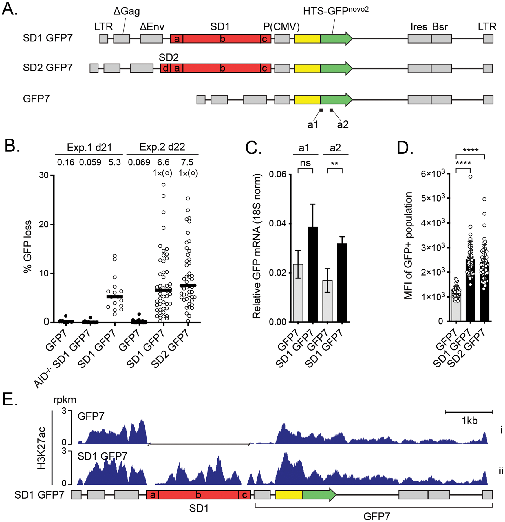
Figure 3.

The effect of DIVAC on the H3K27ac in Ramos cells A. Outline of the lentiviral GFP7 reporter used in Ramos cells with SuperDIVAC1 (SD1 GFP7), SuperDIVAC2 (SD2 GFP7), or without a DIVAC (GFP7) inserted upstream of the transcription unit. The subfragments of the DIVACs are a, human Igλ enhancer; b, chicken Igλ enhancer through 3´ core; c, human Igh intronic enhancer and d, mouse Igλ 3-1 shadow enhancer (32). Locations of amplicons a1 and a2 for RT-qPCR (see C) are indicated.
B. GFP loss of the GFP7 reporters in two independent experiments after 21 and 22 days. SD1 GFP7 was also assayed in AID-deficient Ramos cells (AID−/− SD1 GFP7). Data points outside the y-axis range are in parentheses. Values are medians.
C. Expression of GFP mRNA from integrated SD1 GFP7 reporter assessed using two different RT-qPCR amplicons (a1 and a2) indicated in A. Values are mean +/− SD. Student’s t-test * p<0.05, ** p<0.01, *** p<0.001, **** p<0.0001, ns not significant.
D. Mean fluorescence intensity (MFI) of GFP-positive populations of the reporters in Ramos cells.
E. H3K27ac ChIP-seq of the lentiviral GFP7 reporter with SD1 (SD1 GFP7) and without a DIVAC element (GFP7) in Ramos cells. A gap has been inserted in the track of GFP7 reporter in the place of the SD1 sequence after the alignment. Values are rpkm.
