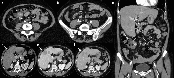
Fig. 3.
Gastrointestinal neuroendocrine carcinoma (NEC) in the terminal ileum. Axial (a, b) and coronal (c) contrast-enhanced CECT images in the arterial phase demonstrate a well-circumscribed enhancing mass (white arrows) of the terminal ileum involving ileocecal valve; in the mesenteric fat near the primary tumour, there is a mesenteric mass (b, c. white circle) with desmpplastic reaction. Arterial phase (d), portal venous phase (e) and equilibrium phase (f) contrast-enhanced MDCT show multiple hypervascular liver metastases (with arrows) with central necrosis

