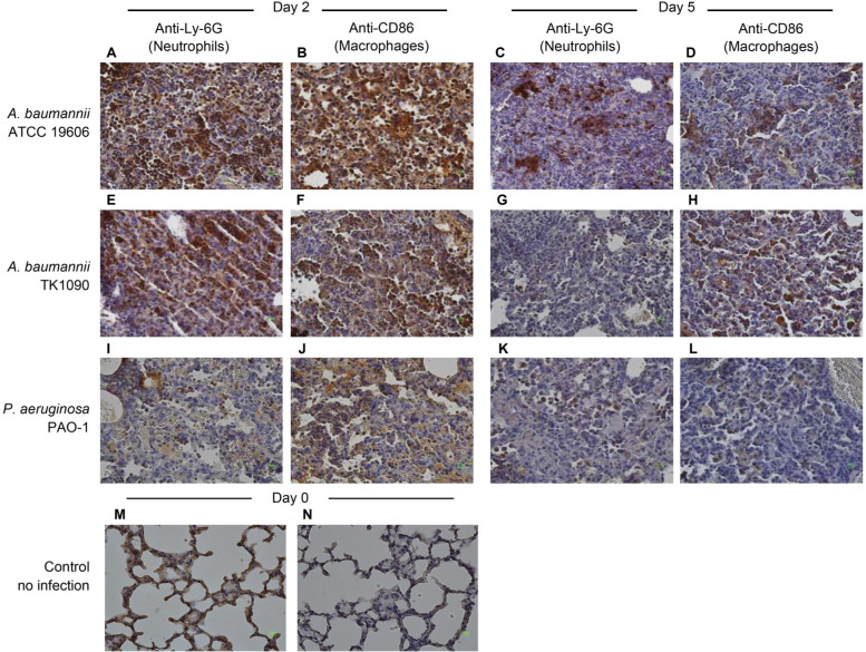
Fig. 4.
Infiltration of neutrophils and macrophages in the lung tissues of Acinetobacter baumannii- and Pseudomonas aeruginosa-infected mice. The lung tissues from C3H/HeN mice infected with ATCC19606 (A, B, C, and D), TK1090 (E, F, G, and H), and PAO-1 (I, J, K, and L) strains and uninfected mice (M and N) are shown. The lung tissues from the mice infected with ATCC 19606 at 1 day (A and B) and 5 days (C and D) post-infection. The lung tissues from the mice infected with TK1090 at 1 day (E and F) and 5 days (G and H) post-infection. The lung tissues from the mice infected with PAO-1 at 1 day (I and J) and 5 days (K and L) post-infection. Neutrophils and macrophages were detected by immunostaining with anti-Ly-6G antibody (A, C, E, G, I, and K) and anti-CD68 antibody (B, D, F, H, J, and L) using diaminobenzidine (DAB) method and restained with hematoxylin, respectively. Photomicrograph images (magnification, 100×).

