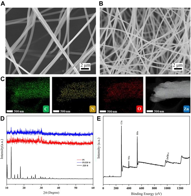
FIGURE 2.
Scanning electron micrographs of the PI-ZIF nanofibers (A) PI and (B) ZIF-8-3/PI. (C) C, N, O, and Zn element distribution of ZIF-8/PI nanofibers. (D) X-ray diffraction patterns of the ZIF-8, PI, and ZIF-8-3/PI nanofiber membranes. (E) X-ray photoelectron spectroscopy images of the ZIF-8-3/PI nanofiber membranes.

