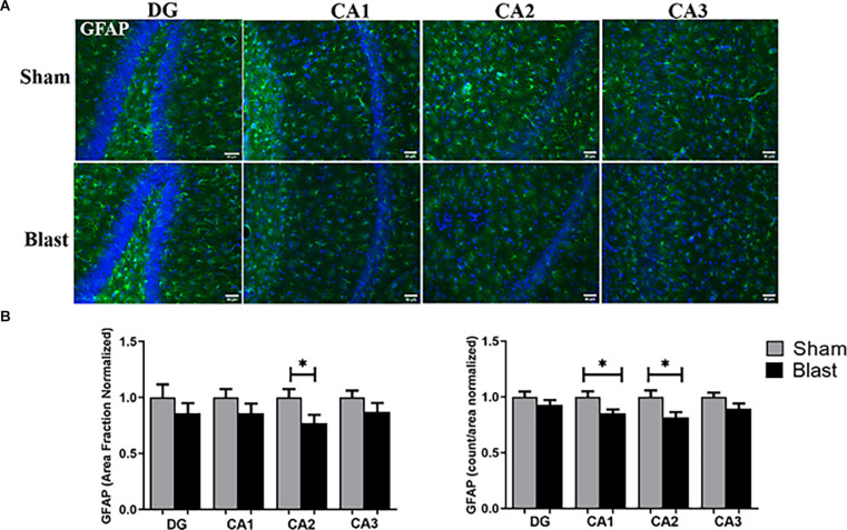
Figure 7.
Decreased levels of GFAP were observed in the hippocampus 52 weeks following blast injury. (A) Representative images of GFAP (green) and DAPI (blue) obtained from the hippocampus region of the brain. Magnification is at 20x and scale bar = 50 μm. (B) Decreased positive GFAP signal (area fraction) was observed in the CA2 sub-region of the hippocampus in blast animals compared to shams. Decreases in the amount of GFAP+ astrocytes were observed in the CA1 and CA2 sub-regions in blast animals. *p < 0.05. Data is represented as Mean ± SEM. DAPI, 6-diamidino-2-phenylindole.

