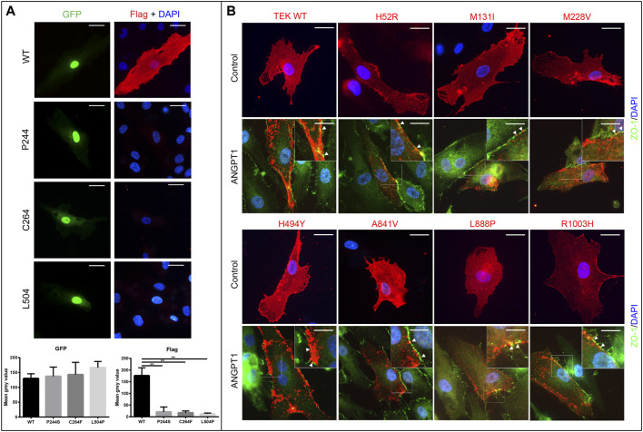
FIGURE 4.
Expression and ligand responsiveness of TEK variants (A). Expression of p.P244S, p.C264F, and p.L504P were greatly decreased in HUVECs. GFP (green) signals indicated cells were successfully transfected. Wild-type Tie2 (anti-Flag, red) was diffusely distributed in cellular membrane of quiescent HUVECs (first row). However, Flag signals for three loss-of-function variants were notably reduced. Scale bars indicate 25 μm. (B) After stimulation by Angiopoietin-1 for 30 min (lower panel), TEK proteins were relocated to cell–cell junctions as indicated by anti-ZO-1 (green). Nuclei were stained by DAPI (blue). Scale bars indicate 25 μm in upper panel and 12.5 μm in lower panel for magnified images. Pictures presented were representative of four biological replicates.

