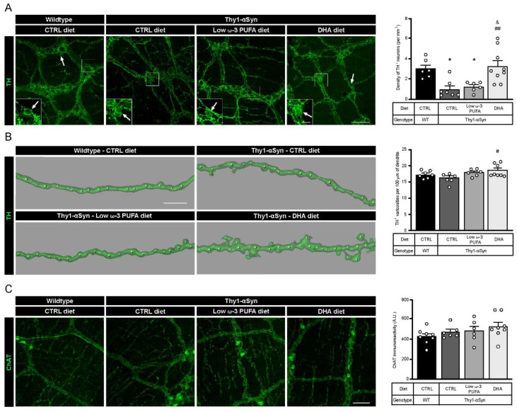
Figure 1.
A DHA-enriched diet restores dopaminergic neuron integrity. (A) Photomicrographs of enteric TH+ neurons (green) and their cell body, as indicated by the white arrow. They show a lower number of dopaminergic cell bodies in the myenteric plexus in the Thy1-αSyn model compared with the wild-type littermates. However, a DHA-enriched diet protected against the decrease in dopaminergic neuron level (one-way analysis of variance (ANOVA): p = 0.0009). Scale bar = 60 µm; Close-up scale bar = 15 µm. (B) Three dimensional images of TH+ dendrites (green) and varicosities (white dots) show an increase of varicosities per 100 μm of dendrites in transgenic mice fed with DHA-enriched diet compared to control diet. No difference was observed between the Thy1-αSyn mice and the wild-type littermates (one-way ANOVA: p = 0.0396). Scale bar = 10 μm. (C) Photomicrographs and immunoreactive quantification of ChAT (green) shows no difference between models or among dietary groups (one-way ANOVA: p = 0.2987). Scale bar = 60 μm. Tukey’s post-hoc tests: * p < 0.05 compared with wild-type mice; # p < 0.05, ## p < 0.01 compared with Thy1-αSyn on control diet; & p < 0.05 compared with Thy1-αSyn on low ω-3 PUFA diet. Abbreviations: A.U., arbitrary unit; ChAT, choline acetyltransferase; CTRL, control; DHA, docosahexaenoic acid; TH, tyrosine hydroxylase; WT, wild-type.

