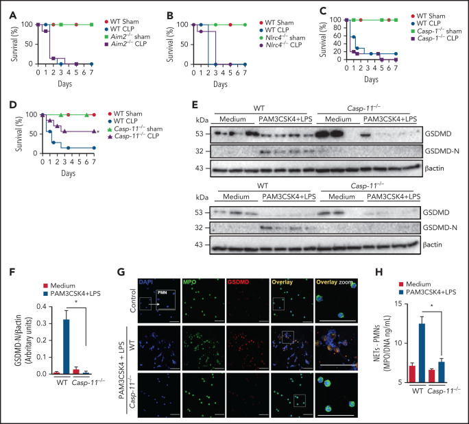
Figure 3.
Caspase-11 contributes to NET release through gasdermin D. (A-D) The survival of WT, Aim2−/−, Nlrc4−/−, Casp1−/−, and Casp11−/− mice subjected or not to CLP was followed for 7 days. (E) Bone marrow neutrophils from WT and Casp11−/− mice were primed with PAM3CSK4 (1 μg/mL) for 4 hours and then transfected with ultrapure LPS (10 μg/mL) for 4 hours. The cell lysates were harvested for immunoblot analysis of GSDMD total and its cleaved fraction (GSDMD-N). Each band represents 1 different animal. Actin (β-actin) was used as a loading control. (F) Quantification of GSDMD-N relative to β-actin. Bone marrow neutrophils from WT and Casp11−/− mice were primed with PAM3CSK4 (1 µg/mL) for 4 hours and then transfected with ultrapure LPS (10 µg/mL) for 4 hours. (G) Representative fluorescence images of NETs stained for DNA (DAPI, blue), myeloperoxidase (MPO, green), and the gasdermin D cleaved fraction (GSDMD, red) are shown. Scale bar, 50 µm at ×630 magnification. (H) The concentration of MPO/DNA-NETs was determined in the culture supernatants. The data are expressed as means ± SEM. *P < .05; H-Mantel-Cox log-rank test (A-D), 1-way ANOVA followed by Tukey’s test (F,H). The data are representative of ≥2 independent experiments, each including 5 to 7 animals per group.

