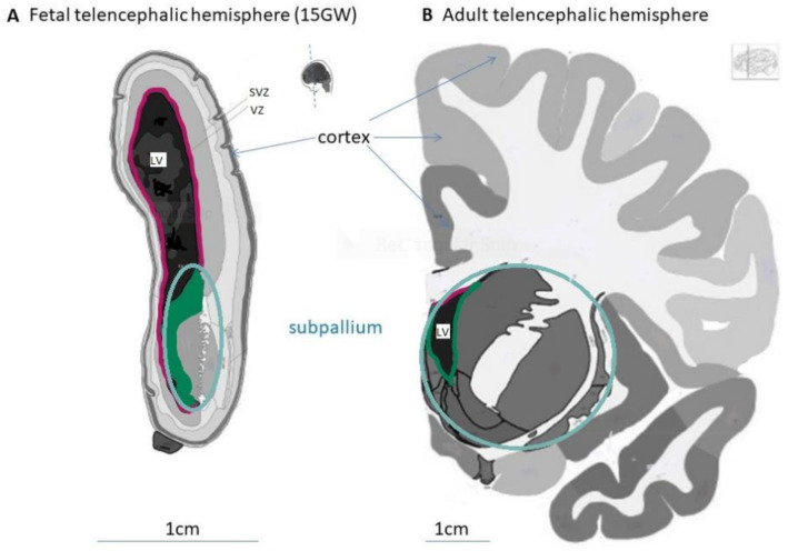
Figure 1.
Morphological structures of the fetal and adult human telencephalon. Coronal sections from fetal (A) and adult (B) telencephalic hemispheres showing the pallium with the cortex, the subpallium and the lateral ventricle (LV). Images modified from the BrainSpan Reference Atlases for 15 gestational week and 34-year-old human brains sectioned at the rostral level (https://atlas.brain-map.org/, accessed on 20 August 2021).

