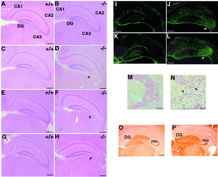
FIG. 4.
Abnormalities in hippocampal CA3 subfields of STAM1−/− mice. (A to H) HE staining of anterior coronal hippocampus sections of mice. Mice were STAM1+/+ (A, C, E, and G) and STAM1−/− (B, D, F, and H) and were 3 weeks old (A and B), 5 weeks old (C and D), 7 weeks old (E and F), and 9 weeks old (G and H). Note the loss of pyramidal cells in the CA3 subfields in STAM1−/− mice (arrowheads). DG, dentate gyrus. (I to L) Anti-GFAP antibody staining. Brain sections derived from STAM1+/+ and STAM1−/− mice were immunostained with anti-GFAP antibody. Gliosis (arrowheads) in hippocampal CA3 subfields was observed in STAM1−/− mice that were 4 weeks old (J) and 7 weeks old (L) but not in STAM1+/+ mice that were 4 weeks old (I) and 7 weeks old (K). (M and N) TUNEL staining of hippocampal CA3 subfields. Hippocampus sections of 4-week-old STAM1+/+ and STAM1−/− mice were stained for TUNEL. Arrowheads indicate positive staining for TUNEL. (O and P) Calbindin immunostaining. The sections were prepared from the brains of 9-week-old STAM1+/+ and STAM1−/− mice. DG and mo, dentate gyrus and mossy fibers, respectively. Bars, 0.4 mm.

