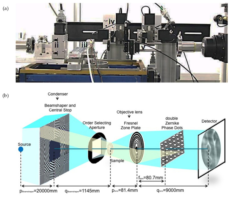
Figure 3.
(a) Photo of the phase-contrast nanotomography setup at TOMCAT, showing (i) the end of the X-ray fly-tube, (ii) the condenser, (iii) the environmental chamber housing the wood sample, (iv) the Fresnel zone plate and (v) the Zernike phase dots. The camera is located at 9 m of distance from the (iv) and is not shown in the picture. (b) Schematic representation of the nanotomographic microscopy, with bordered pits imaged on the detector plane.

