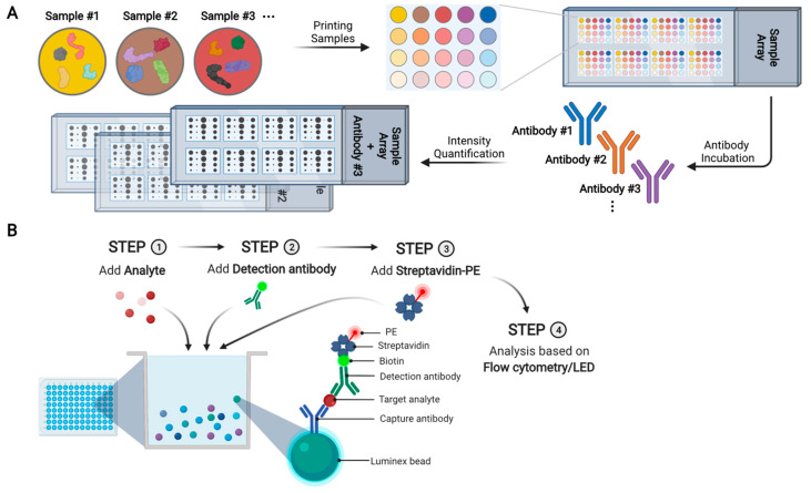
Figure 8.
Schematic overview of reverse phase protein array (RPPA) and Luminex microsphere bead capture. (A) Schematic of RPPA. Each spot contains a single sample and each array is probed with one specific antibody. The bound antibody can be quantified (either directly or indirectly) by fluorescent, colorimetric, or chemiluminescent assays. (B) Schematic of Luminex microsphere bead capture assay. Analyte-specific capture antibodies are immobilized on superparamagnetic microsphere beads that are color-coded. After incubating a test sample with antibody-coated microsphere beads, target proteins are captured. Biotinylated detection antibodies specific to the target proteins are added, leading to the formation of an antibody–antigen sandwich. Phycoerythrin (PE)-conjugated streptavidin is added, so that the protein amount can be quantified based on the intensities of PE-derived signal.

