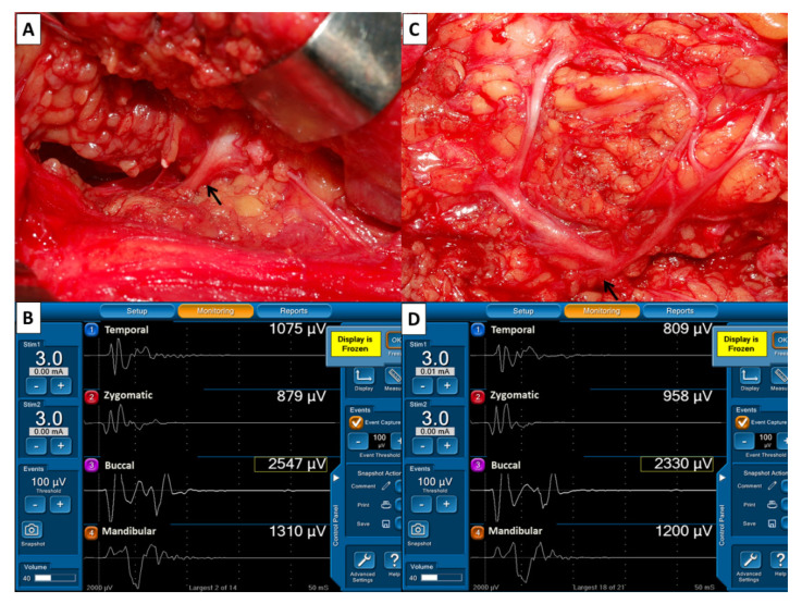
Figure 2.
Evaluation of facial nerve (FN) function before and after dissection of FN branches. (A) After identification of the main trunk of the FN (↑), a stimulus current of 3 mA was applied. (B) The EMG amplitudes of four elicited signals were designated F1 signals and used as reference values for FN function. (C) After dissection of FN branches and resection of the parotid tumor, the main trunk of FN (↑) was stimulated with the same stimulus current, and (D) the elicited EMG signals were designated F2 signals. On each channel, EMG amplitudes were compared between F2 and F1 signals. This showed 25% of decreased amplitude on channel 1 (809/1075 µV), 11% of increased amplitude on channel 2 (958/879 µV), unchanged amplitude on channel 3 (2330/2547 µV) and channel 4 (1200/1310 µV).

