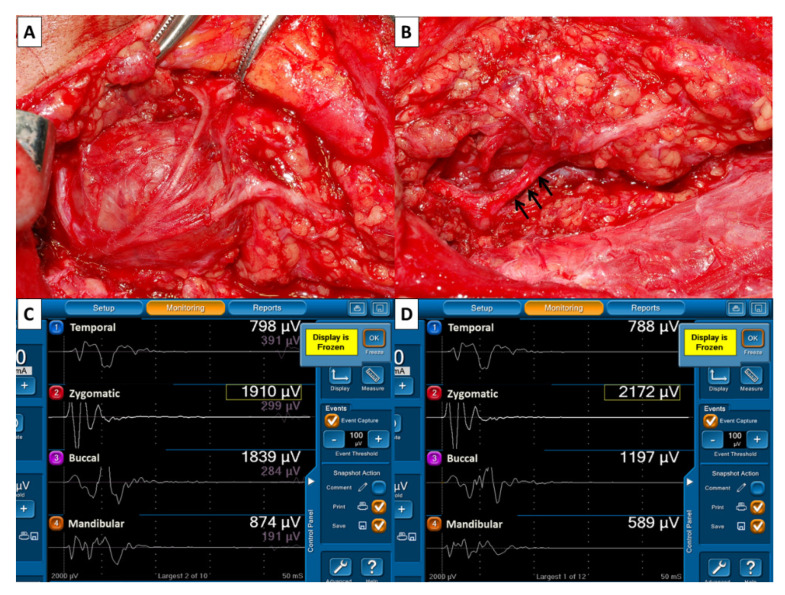
Figure 5.
(A) The FN branches adhered to the deep lobe parotid tumor. (B) A segment of the inferior cervicofacial branch (↑) had a red swollen appearance after tumor resection. Comparison of EMG amplitudes of F2 (C) and F1 (D) signals revealed unchanged amplitude on channel 1 (788/798 µV) and channel 2 (2172/1970 µV), 35% (1197/1839 µV) and 33% (589/874 µV) of decreased amplitude on channel 3 and 4. The patient had normal facial expression after surgery.

