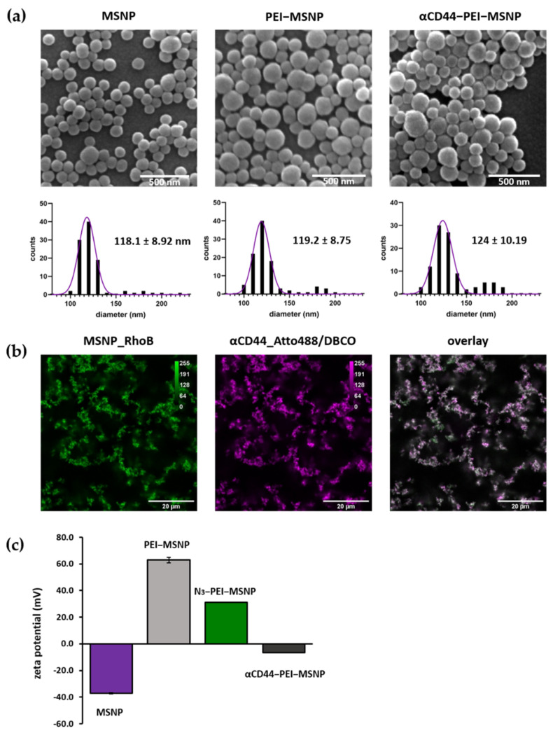
Figure 3.
Characterization of bare (MSNPs), PEI-coated (PEI-MSNPs), Azide-functionalized (N3-PEI-MSNPs), and CD44-conjugated (αCD44-PEI-MSNPs) mesoporous silica nanoparticles. (a) Representative SEM images and size distribution of the imaged particles. Values shown as mean ± SD. Scale bar is 500 nm. (b) Confocal fluorescence images of αCD44-PEI-MSNPs in which MSNPs were loaded with RhoB (first panel, green), while an Atto488 (and DBCO) label was conjugated to the CD44 antibody (second panel, magenta). An overlay is displayed in the third panel. Scale bar is 20 µm. (c) Zeta-potential measurements given as mean ± SD.

