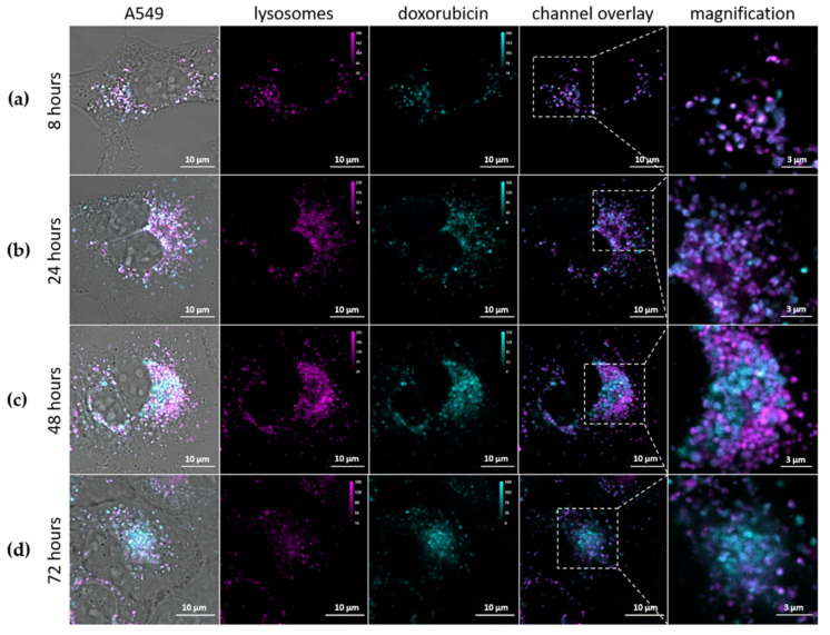
Figure 6.
Intracellular release of Dox (cyan) from αCD44-PEI-MSNPs_Dox with respect to the lysosomes (magenta) over time. A549 cells were incubated with αCD44-PEI-MSNPs_Dox (final concentration of 50 μg/mL) for (a) 8 h, (b) 24 h, (c) 48 h and (d) 72 h. The first column shows a complete merge including the transmission image, also displaying the cell contours. In the second and third columns, the lysosomes and Dox are depicted, in pink and cyan, respectively. In the fourth and fifth columns, a channel overlay and respective magnification are shown. Scale bar is 10 μm in the main images and 3 μm in the magnified images.

