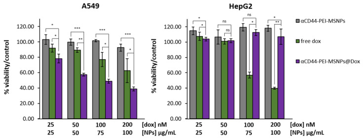
Figure 7.
Viability of A549 and HepG2 cells after 72-h incubation with different concentrations of free Dox, Dox-loaded αCD44-PEI-MSNPs, and empty αCD44-PEI-MSNPs. A549 and HepG2 cells that were not incubated with particles or drug were used as a control and represent 100% viability (data not shown). Dox concentrations are expressed in nM while nanoparticle concentrations are in µg/mL. Estimated Dox concentration loaded in the nanoparticles was 50 μM. Error bars indicate ± SD, with ns meaning not significant; * (p < 0.05), ** (p < 0.01), and *** (p < 0.001), n = 4.

