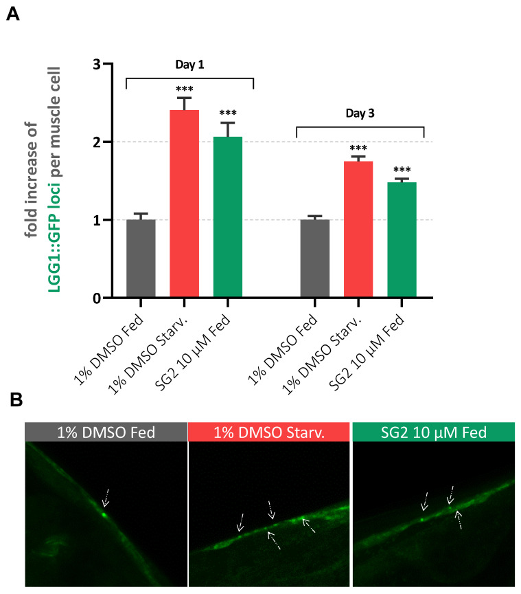
Figure 3.
SG2 enhances autophagy in a C. elegans model of AD. (A) GFP structures were counted in 3 different conditions (Fed: 1% DMSO; Starved: 1% DMSO and 6 h of starvation; Treated: 10 µM SG2 in 1% DMSO) of approximately 25–30 worms from two different biological experiments. Bars represent average number of GFP structures normalized to control (Fed) + SEM. For statistical tests, One-Way ANOVA was used p < 0.001 ***. (B) Representative pictures of big muscles cells presenting LGG-1:GFP puncta with white arrows highlighting LGG-1::GFP structures.

