Figure 1.
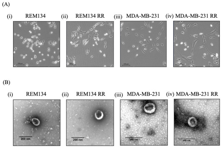
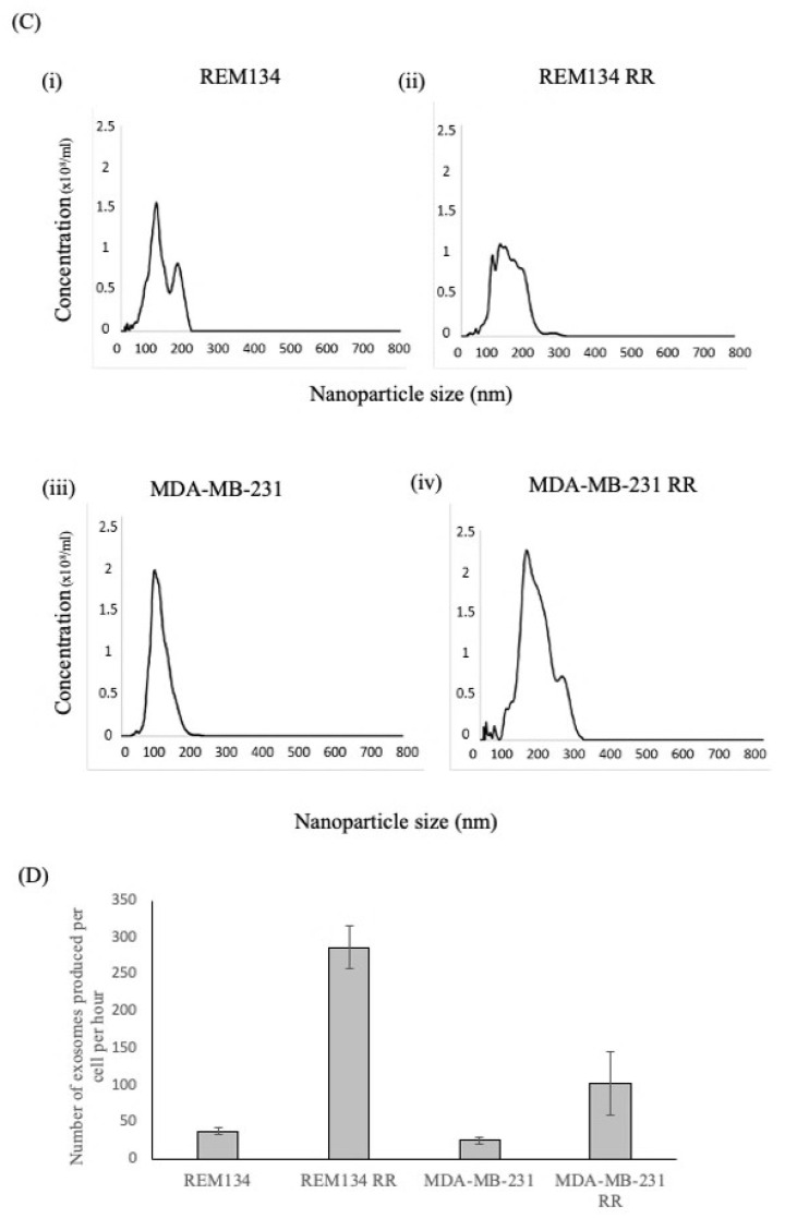
Isolation of exosomes from canine and human breast cancer cell lines and their derived isogenic RR counterparts. (A) Cell morphology of (i) REM134, (ii) REM134 RR, (iii) MDA-MB-231 and (iv) MDA-MB-231 RR cells. Scale bar represents 100 μm. (B) Visualisation, using TEM, of exosomes isolated from (i) REM134, (ii) REM134 RR, (iii) MDA-MB-231 and (iv) MDA-MB-231 RR cells. Scale bar represents 200 nm. Characterisation of exosomes using NTA to measure (C) particle distribution from (i) REM134 cells, (ii) REM134 RR, (iii) MDA-MB-231 and (iv) MDA-MB-231 RR and (D) rate of exosome production per cell per hour. Data are representative of three independent experiments.


