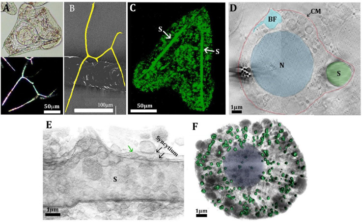
Figure 2.
Mineral deposition process in sea urchin larvae. (A) Light micrographs of a live larva of the sea urchin Paracentrotus lividus taken 41 h after fertilization (hpf) (top image); image at the bottom: the same larva observed under polarized light, showing the crystalline nature of the spicules. (B) SEM micrograph of an isolated spicule from Litechinus pictus larva. The spicule is pseudocolored yellow to facilitate observation. (C) Confocal fluorescence image of a 46hpf larva developed continuously in calcein-labeled seawater. Note that the spicule (S) is fully labeled. Many intracellular vesicles are also fluorescent, indicating uptake by endocytosis of seawater. (D) Cryo-FIB-SEM micrograph of a high-pressure frozen 40hpf larva. The cyan vesicle is open toward the body cavity (blastocoel), which is filled with a seawater-like solution. BF, blastocoel fluid; CM, cell membrane; N, nucleus. (E) Segmentation of cryo-FIB-SEM serial milling and block face imaging stack acquired from 40hpf high-pressure frozen larva. The segmentation shows the syncytium enveloping the spicule. A vesicle (green arrow) is depositing onto the growing spicule. Reprinted and modified with permission from (41). Copyright 2016 Elsevier. (F) Segmentation of cryo-soft X-ray tomography of a PMC taken from 36hpf larva. The colored particles are Ca-rich particles. Cytoplasm and other cellular vesicles and organelles are gray.

