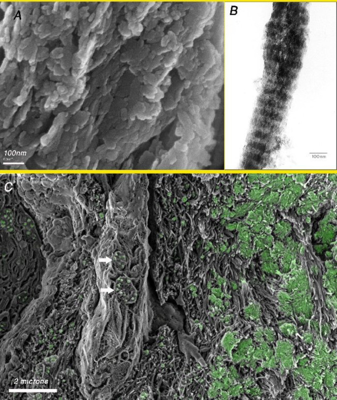
Figure 3.

(A) SEM image of fractured baboon tibia after removal of the organic matrix using sodium hypochlorite. Note the plate-shaped crystals organized in layers. (B) TEM image of an isolated mineralized collagen fibril extracted mechanically from turkey tendon. The banding is due to the presence of more plate-shaped crystals in the gap region of the collagen fibril as compared to the overlap region. (C) Cryo-SEM image of the fracture surface of an embryonic chicken bone showing in green the distribution of mineral (based on the BSE image of the same area). Note the presence of vesicles containing mineral particles inside the cell (arrows).
