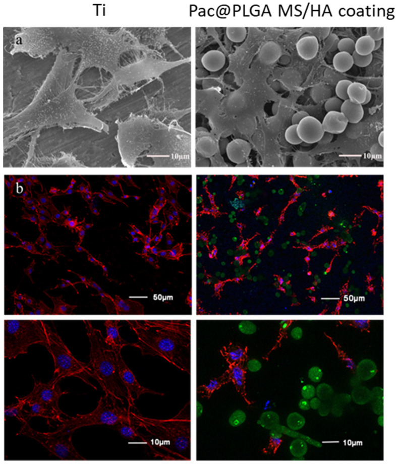
Figure 3.
The adhesion of MC3T3-E1 cells cultured on Ti and Pac@MS/HA coating of Ti for 6 h was observed through SEM and CLSM. (a) SEM images of MC3T3-E1 cells on Ti and Pac@PLGA MS/HA coated Ti surface. (b) Confocal images of MC3T3-E1 cells on Ti and Pac@PLGA MS/HA coated Ti surface. The blue fluorescence is from DAPI in the nucleus; the red fluorescence is from phalloidin in the cell membrane; and the green fluorescence is from Pac@PLGA microspheres.

