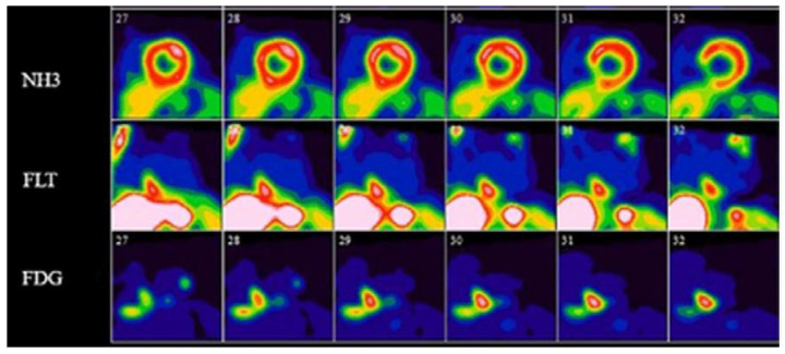
Figure 5.
Short axis 13N-ammonia perfusion, 18F-FDG-, and 18F-FLT-PET images in a patient with active cardiac sarcoidosis. 13N-ammonia perfusion images (top row) show scarring of the inferior ventricular septum, with both FDG- and FLT-PET images showing evidence of active cardiac sarcoidosis in this area. Reprinted from ref. [75].

