Figure 4.
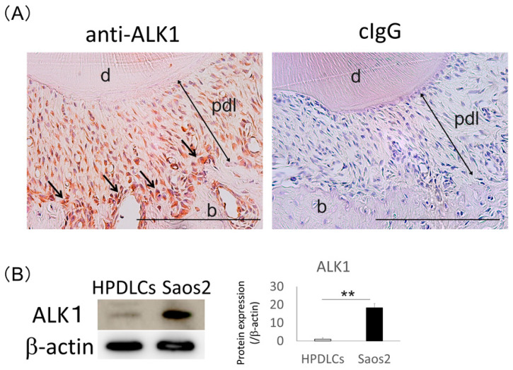
Expression of ALK1 in normal rat periodontal ligament (PDL) tissue and Saos2 cells. (A) Immunohistochemical analysis was performed using an anti-ALK1 antibody in rat PDL tissue. Positivity to an anti-ALK1 antibody was detected in PDL tissue, and ALK1-immunoreactive cells were localized in the osteoblastic layer (arrows). Control IgG was used for a negative control. Hematoxylin was used for the staining of nuclei. Experiments were performed in duplicate. b, alveolar bone; d, dentin; pdl, periodontal ligament. Bars = 500 µm. (B) Western blotting analysis was performed to detect expression of ALK1 in human PDL cells (HPDLCs) and Saos2 cells. The expression levels of this protein were normalized against β-actin expression and were quantified. The expression level of ALK1 was higher in Saos2 cells than in HPDLCs. Values are the means ± SD from three independent experiments. ** p < 0.01.

