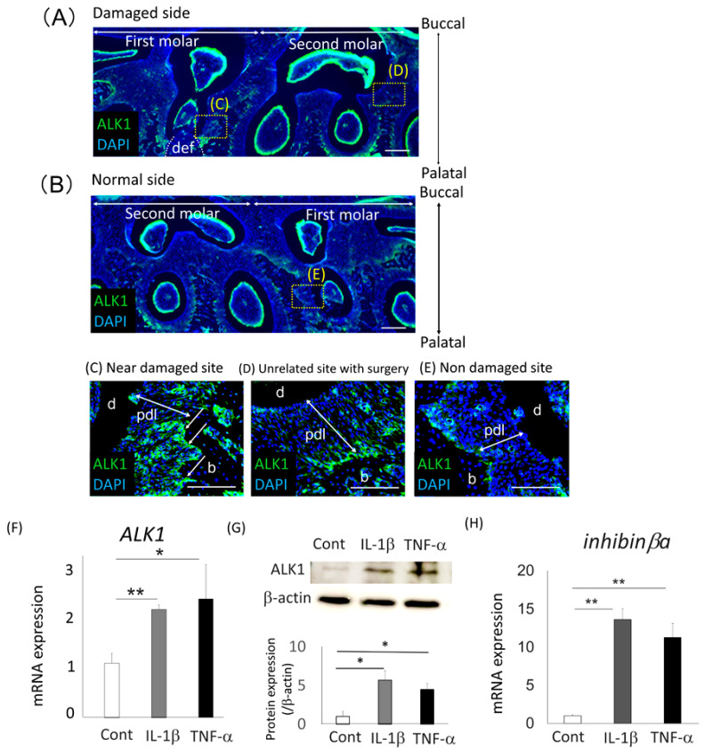
Figure 5.
Expression of ALK1 around damaged rat periodontal tissue and in IL-1β- or TNF-α-treated Saos2 cells (A–E) Immunofluorescent analysis of ALK1 expression (green) in a surgically damaged side (A) and a normal side (B) of rat periodontal tissue was performed. The tissue samples were collected 3 days after surgery, and horizontal sections of the first and second molars were prepared from the rat maxilla. Panels C-E show higher magnification views of panels A and B. More intense staining was detectable around the osteoblastic layer in the defect site (def) (C), compared with normal periodontal tissue from the second molar in the damaged site (D) or the first molar from the normal site (E) (arrows). Nuclei were stained with 4,6-diamidino-2-phenylindole (DAPI; blue). Bars = 100 µm. b, alveolar bone; d, dentin; pdl, periodontal ligament. (F,H) Gene expression of ALK1 (F) and inhibinβa (H) in Saos2 cells treated with IL-1β or TNF-α for 24 h was analyzed using quantitative RT-PCR. Untreated cells were used as the control. Normalization of gene expression was performed against β-actin expression, and the gene expression levels were shown as the fold increase of the control. Values were the means ± SD from three independent experiments. ** p < 0.01, * p < 0.05. (G) Western blotting analysis was performed to detect expression of ALK1 in Saos2 cells. Normalization of protein expression was performed against β-actin expression and the expression level of this protein was quantified. The results were shown as the fold increase of the control. Values are the means ± SD from three independent experiments. * p < 0.05.

